

Oral Cancer
Although this topic is scary, this article may just save your life
(Continued)
 |
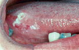 |
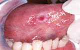
| Figure 1: Early cancer (squamous cell carcinoma) that was first thought to be a harmless sore (ulcer) caused by biting the tongue. | Figure 2: Early cancer (squamous cell carcinoma) that was first mistaken for a harmless white patch (benign leukoplakia). |
 |
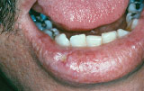 |
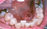
| Figure 3: Early cancer (squamous cell carcinoma) of the floor of the mouth was noticed for 2 weeks and at first was thought to be a canker sore. | Figure 4: Early cancer (squamous cell carcinoma) of the lip was noticed for 1 month and was at first thought to be a “cold sore.” |
| Photos provided by Dr. Sol Silverman, Jr. | |
Stick Out Your Tongue
The tongue, particularly the sides are the most common sites for oral cancer [Figure 1 and 2], with the floor of the mouth (under the tongue) coming in second [Figure 3]. Lip cancers mostly affect the lower lip [Figure 4] and frequently there is a history of chronic sun exposure and preceding damage, which shows up as scaling and crusting at the site. The thing to remember here is that recurring ulcers in the lip area can also be mistaken for cold sores. Since the tongue has a rich blood supply and lymphatic drainage (the lymphatic system is a major component of our immune protection system) 30% of cancers have spread or metastasized by the time they are diagnosed. That's a frightening fact. Now let's take that fact a step further — up to 15% of people diagnosed with oral cancer are normally found to have a second primary cancer.
However, when detected early while a lesion is small, survival rate exceeds 80%. Bear in mind, early detection is key. If you notice any unusual lesions (sores or ulcers), or color changes (white or red patches), anywhere in your mouth that do not heal within two-three weeks get to your dentist or physician as soon as possible.
The Oral Cancer Exam
An oral cancer examination should be part of your dental check-up or regular cleaning appointment. The oral cancer examination consists of the following:
- A visual inspection of your face, neck, lips, and mouth looking for any signs of cancer (such as red and/or white patches).
- Your dentist will feel the floor of the mouth, sides of the neck, glands etc. for any lumps that may suggest cancer.
- Using gauze your dentist will gently pull your tongue from side to side as well as examine the underside of it.
- Your dentist will also ask you to say “Ahh” and will then place an instrument on top of your tongue to examine the back of your throat.
Diagnosis Can Be Complicated
Earlier we talked about the fact that oral cancers are most often detected when they are at a late stage, with early diagnosis only taking place in about one third of the cases. Unfortunately, recognition is complicated. Why? Because the early signs can mimic harmless sores that occur in the mouth such as canker sores, minor infections, or irritations that occur from biting or even certain foods. When we're given a proper oral cancer exam which includes the oropharynx, the health care professional will feel the neck for lumps; inspect the lips and all inside surfaces of the mouth, including the tonsils at the back of the throat.
Further, we must remember that oral cancers can occur on any surface that lines the mouth and throat, with tongue being the most common site. These changes — as I mentioned earlier — can appear as white or red patches, ulcers and lumps that may or may not be associated with any discomfort or pain.
An appropriately trained dentist should evaluate any such changes that persist for more than two-three weeks. Definitive diagnosis requires the microscopic examination of a piece of the lesion (tissue biopsy). This is a procedure usually carried out with local anesthesia, numbing of the involved site with the removal of a sample or all of the abnormal tissue, if small enough. The tissue specimen is then sent to the lab for analysis where it undergoes microscopic evaluation for a more thorough diagnosis.

